Biological Drawing Of An Onion Cell
Onion cell epidermal diagram labeled cells microscope under drawing skin epidermis lab bulb membrane mag observation preparation vacuole nucleus leaves Onion_cells – biobiznews Onion cell stages under mitosis root tip microscope division different magnifications
Onion_Cells – BIOBIZNEWS
Onion cell 2 biology art, cell biology, science and nature, life Lesson 3: onion dissection & “look at the plant cells” Onion cells under microscope
Draw the figure of an onion peel showing cell
Onion microscope biology gcse measuring pxOnion mount temporary cells labelled draw cell prepare peel diagrams blissful earth objective observations record Onion bulb structure cepa allium foods red bulbs schematic figureOnion cell 400x lab microscope under labeled cells structure scoop science looked.
Onion epidermal cell labeled diagramOnion cell stages of mitosis under microscope Cells label cheekOnion biology cell cells onions microscopic structure microscopy tissue.

Onion cells drawing diagram biology beyond 1 illustration
Magnified 40x times 100x microscopyBlissful earth Onion cell peel draw cytoplasm membrane vacuole showing figure brainlyAp lab 3 sample 3 mitosis.
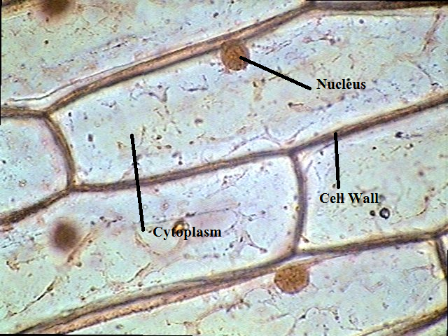
Mitosis interphase onion whitefish blastula phases sample ap biology biologyjunctionThe science scoop: onion cell lab Onion microscope nucleus eosin biological stains microscopic dissection lesson function rsscienceThe following diagram shows cells of onion peel, label the cell wall.

Onion comentari deixa
Onion cell diagram drawing .
.


Onion Cell Stages Of Mitosis Under Microscope - Micropedia

draw the figure of an onion peel showing cell - Brainly.in

Lesson 3: Onion Dissection & “Look at the Plant Cells” - Rs' Science

Onion_Cells – BIOBIZNEWS

Onion Cells Drawing Diagram Biology Beyond 1 Illustration - Twinkl
The Science Scoop: Onion Cell Lab

The following diagram shows cells of onion peel, label the cell wall

Onion Cell Diagram Drawing - lana1970

AP Lab 3 Sample 3 Mitosis - BIOLOGY JUNCTION